Blog:EMG vs. MRI: Differences, Uses, and Which Test Is Right for Nerve Pain
EMG vs. MRI: Differences, Uses, and Which Test Is Right for Nerve Pain
When numbness, tingling, weakness, or unexplained pain starts interfering with everyday life, it is natural to wonder which test will reveal the problem. Two tests that often come up are electromyography (EMG) and magnetic resonance imaging (MRI).
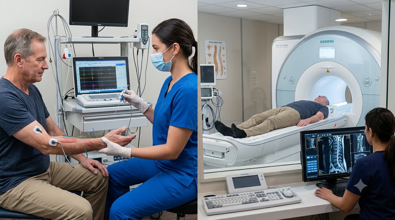
Although both can help doctors investigate neurological symptoms, they do very different jobs. An EMG evaluates the electrical activity of muscles and the function of the nerves that control them. An MRI creates detailed images of tissues and structures inside the body. Neither test is automatically better than the other. The right choice depends on what a neurologist is trying to find.
That distinction becomes particularly important with symptoms such as a suspected pinched nerve, back pain, sciatica, weakness, or persistent tingling. In some situations, an EMG or nerve conduction study may provide information that an MRI cannot. In others, an MRI may be more useful because the concern is a structural problem. Sometimes both tests are appropriate because they answer different questions.
EMG vs. MRI: What Is the Main Difference?
The simplest way to understand the difference is to think of function versus structure.
An EMG looks at how nerves and muscles are working. During the test, electrical activity in selected muscles is recorded, usually with a small needle electrode. A nerve conduction study is commonly performed along with the EMG. It uses electrodes on the skin to measure how quickly and effectively electrical signals travel through nerves.
An MRI, on the other hand, is an imaging test. It uses a strong magnetic field and radio waves to produce detailed pictures of tissues and structures. Depending on the area being examined, an MRI can help identify problems involving the spine, discs, nerves, brain, muscles, or other soft tissues.
So if a doctor is asking, “Is this nerve functioning normally?”, an EMG may be particularly useful.
If the question is, “Is there a structural problem that could be affecting this nerve?”, an MRI may provide the more relevant information.
What Does an EMG Show?
An EMG is used to assess the health and function of muscles and the nerves that control them. It can help investigate symptoms such as muscle weakness, numbness, tingling, cramping, and certain types of limb pain.
The test can provide useful information about conditions involving peripheral nerves, including some forms of neuropathy, carpal tunnel syndrome, and nerve injuries. It can also help evaluate whether a nerve root may be affected, such as with some cases of a herniated disc.
One important advantage is that EMG testing provides information about nerve and muscle function, rather than simply showing what an anatomical structure looks like.
For example, someone may have persistent tingling and weakness in one hand. An MRI might show structures in the neck that could potentially affect a nerve. An EMG and nerve conduction study can provide additional information about whether the nerve is actually conducting signals normally and where dysfunction may be occurring.
Bansal Neuro's EMG service notes that testing can help neurologists evaluate conditions including neuropathy, pinched or damaged nerves, carpal tunnel syndrome, and other neuromuscular conditions.
What Does an MRI Show?
MRI answers a different question.
Instead of measuring electrical signals, it produces detailed images of the body's internal structures. For neurological complaints, doctors may use MRI to examine areas such as the brain or spine and look for structural abnormalities.
For example, if a person has back pain with symptoms traveling into the leg, an MRI may help identify a herniated disc or another structural problem that could be pressing on a nerve. MRI is particularly useful for visualizing soft tissues of the spine and can help show the location of a herniated disc and which nerves may be affected.
MRI can therefore be valuable when the suspected cause of symptoms involves anatomy that needs to be visualized.
But an MRI image does not necessarily tell the entire story. A scan can show an abnormality without proving that the abnormality is responsible for someone's symptoms. That is one reason a neurological examination and, when appropriate, functional testing such as EMG may also be considered.
EMG or MRI for a Pinched Nerve?
This is one of the most common situations in which people ask about EMG vs. MRI.
The answer depends on what the doctor needs to determine.
An MRI can show whether there is a structural reason for nerve compression. For example, it may reveal a herniated disc or another abnormality around the spine.
An EMG can help determine whether there is evidence of nerve dysfunction affecting the muscles. Nerve conduction testing can also provide information about how electrical signals are traveling along the nerve.
Consider a patient with pain, numbness, and weakness extending from the lower back into the leg. If the concern is a disc problem pressing on a nerve root, an MRI may help identify the structural cause. If the neurologist also needs to determine whether a nerve is functioning abnormally or distinguish between different possible sources of nerve dysfunction, EMG and nerve conduction testing may add important information.
That is why there is not a universal answer to “Is EMG or MRI better for a pinched nerve?” The tests provide different kinds of information.
EMG vs. MRI for Back Pain and Sciatica
Back pain does not always come from the same source, and pain traveling down an arm or leg can have several possible explanations.
An MRI may be considered when a structural problem involving the spine is suspected. It can provide detailed images of discs and other soft tissues and may help identify conditions such as a herniated disc.
EMG and nerve conduction studies may be useful when the clinician needs more information about nerve or muscle function. An EMG can help identify whether particular nerves are damaged or whether muscle abnormalities may be contributing to symptoms.
This distinction matters because pain does not always equal nerve damage. A person's symptoms, neurological examination, medical history, and test results need to be considered together rather than interpreting one scan or test in isolation.
Can You Need Both an EMG and an MRI?
Yes.
An EMG and MRI are not competing tests. In some cases, using both can give a more complete picture.
Imagine an MRI showing a narrowing in the spine near a nerve. The image tells the physician that a structural abnormality exists. An EMG may provide additional information about whether the nerve and muscles show signs of functional impairment.
The opposite situation can also be informative. A patient may have symptoms suggesting a peripheral nerve problem, while an MRI does not provide an obvious explanation. Functional testing may then help investigate how the nerves are conducting signals.
Mayo Clinic lists both EMG and MRI among diagnostic tests that may be used when evaluating peripheral nerve injuries, with each test providing different information.
The goal is not to order every possible test. It is to select the tests that answer the clinical questions raised by the patient's symptoms and examination.
What Is an EMG Appointment Like?
People sometimes avoid an EMG because they are unsure what the test involves.
An EMG typically involves small electrodes and, for the needle portion of the examination, a thin needle electrode placed into selected muscles to record electrical activity. Nerve conduction testing may be performed at the same appointment using electrodes placed on the skin.
Bansal Neuro states that its EMG appointments generally take about 30 to 60 minutes, depending on the areas being evaluated, and that patients can return to normal activities afterward.
The experience can vary from person to person, but the purpose of the test is straightforward: to gather objective information about how the nerves and muscles are functioning.
What Is an MRI Appointment Like?
An MRI is performed using a scanner that creates detailed images of the body. Unlike an EMG, it does not measure electrical activity in nerves or muscles.
The experience is also different. During an MRI, the patient typically needs to remain still while images are acquired. The length and preparation can vary depending on the body part being examined and whether contrast is needed.
MRI also has considerations that an EMG does not. Because it uses a powerful magnetic field, patients need to tell the imaging team about certain implants, devices, or metal in the body before the scan.
Which Test Is Right for You?
There is no reliable way to choose between EMG and MRI based on one symptom alone. A neurologist may consider factors such as:
Where the symptoms occur
Whether there is numbness, weakness, pain, or tingling
How long the symptoms have been present
Findings from a neurological examination
Whether a spinal or other structural problem is suspected
Whether the concern involves a peripheral nerve, nerve root, or muscle
Whether previous testing has already identified a possible cause
The important question is not simply “Should I get an EMG or MRI?” It is “What does my doctor need to find out?”
That question helps explain why two people with similar symptoms may be sent for different tests.
When Should You Talk to a Neurologist?
Persistent numbness, tingling, unexplained weakness, nerve pain, or muscle symptoms deserve medical evaluation, particularly when they interfere with normal activities or continue without an obvious explanation.
A neurologist can begin with the history and neurological examination before deciding whether further testing is appropriate. Depending on the suspected condition, that may include EMG and nerve conduction studies, MRI, or another diagnostic approach.
Bansal Neuro, operating as A Ray of Hope, provides EMG testing through its neurology practice and lists locations in Libertyville, Illinois; Kenosha, Wisconsin; and Phoenix, Arizona.
The most useful test is ultimately the one that answers the clinical question at hand. An MRI can show what is happening structurally. An EMG can show how nerves and muscles are functioning. Understanding that difference makes the choice between them much less confusing, and in some cases, it explains why a neurologist may recommend both.
© 2026 A Ray of Hope Neurology and Psychiatry. All rights Reserved - Accessibility Statement - Privacy Policy - Sitemap
Managed and Designed by 